
C4: Optofluidic laser
Optofluidic lasing can be a powerful sensing technique for many biochemical processes which occur in an aqueous environment. It combines laser technology and microfluidics, and detects slight changes in environmental conditions which influence the generation of laser light.
A laser generally consists of three main parts:
- Pump source, which provides energy to the system,
- Active medium, in which the amplification of light actually takes place,
- Resonator, in which laser light circulates and thus builds up even more.
Microlasers exist in a variety of geometries, including whispering-gallery resonators, Fabry Perot resonators, photonic crystal resonators and others.[1] In the AG Hunger, we implement fibre-based Fabry-Perot cavities, which consist of two highly reflective concave mirrors fabricated directly on the ends of optical fibres, such that the centre wavelength of the stop band of the mirrors corresponds to the desired laser output. [2]
Pumping in an optofluidic laser is typically performed optically, at a wavelength chosen to match the absorption band of the gain medium. In the laser built in our group, off-resonant longitudinal pumping is done by coupling in pump light along the resonator axis via the resonator fibres themselves.
Following excitation by a pump source, atoms or molecules in the gain medium decay by spontaneously emitting fluorescence photons. In the presence of a resonator, though, these photons circulate within the medium and cause stimulated emission. The threshold to lasing is crossed when the gain exceeds the loss in the cavity.
Laser output characteristics, such as the lasing threshold, are highly sensitive to the conditions in the gain medium. As a result, monitoring the laser output can enable the measurement of small changes in the microfluidic system, such as active medium concentration, temperature and refractive index. [3]
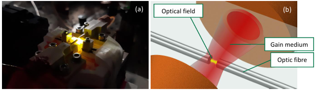
Figure 1 (a) shows the operation of a microlaser using Rhodamine 6G as the gain medium and laser emission around 594 nm, pumped with green light (532 nm). The main components of the system are illustrated in Figure 1 (b).
In the context of CRC 1573, it is of interest to investigate europium-based inorganic-organic hybrid nanoparticles as the lasing medium. The organic ligands shield the Eu3+ cation from the aqueous environment in order to prevent quenching, and also transfer energy to the cation via the antenna effect, allowing for broadband excitation with ultraviolet light. [4] As mentioned earlier, the laser output is strongly dependent on conditions in the gain medium, and therefore, a europium-based optofluidic laser could enable sensing of biochemical processes such as the metabolism of lanthanide-dependent bacteria.
References
[1] H. Zhang et al., Optofluidic lasers and their applications in biochemical sensing. Lab on a Chip 2023, 23, 2959-2989.
[2] M. Gerdan, Optofluidic microlaser based on molecular complexes with the prospective of biochemical process examination. Master’s thesis, KIT (2024).
[3] X. Fan et al., The potential of optofluidic biolasers. Nature Methods 2014, 11, 141-147.
[4] A. Kuzmanoski et al., Tb2(bpdc)3 and Eu2(bpdc)3 nanoparticles and their luminescence. Zeitschrift für Naturforschung 2014, 69b, 248-254.
